The Tangled Tree is also an Optimizing Network – Telling the Story of Molecular Convergent Evolution

The Tangled Tree, David Quammen (2019)

I have just read David Quammen’s The Tangled Tree: A Radical New History of Life (2019). It is a beautifully written book on molecular phylogenetics. Quammen has written over a dozen books on the life sciences, and he is a great storyteller and science journalist.

I recommend this book, with one serious reservation. It describes a purely evolutionary view of molecular phylogenetics. Quammen unfortunately entirely ignores convergent evolution, and thus never allows the reader to consider its implications for universal development. He also does not discuss evo-devo biology. If he had, he might have recognized just how constraining accretive processes of biological development must be on all macrobiological evolutionary change.

Consider the fact that all complex animals, including humans, share almost all the same basic developmental regulatory machinery found in much simpler organisms than us. Like a tree that grows outward from a central trunk, we can’t update our developmental code as we grow more complex. We can only add to that code, progressively limiting our morphological and functional options in evolution. Constraining factors like accretive regulatory development and convergent evolution are physical realities we must recognize if we are to understand long-range macrobiological change on Earthlike planets.

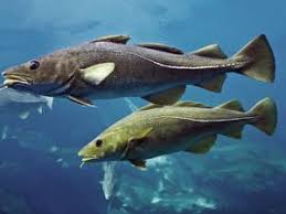
Convergent evolution in antifreeze proteins in Arctic and Antarctic fish.

Scientists have been researching the molecular phylogenetics of convergent evolution since the 1990s, when evo-devo biology first became a formal subdiscipline. For example, we’ve known since 1997 that antifreeze proteins evolved via two clearly independent genetic means in Northern and Southern polar fish, to prevent ice crystal formation.

As our science and simulation advance, I think we will discover a vast number of developmental portals, uniquely adaptive and accelerative attractors on the road to competitive complexification that must be discovered via evolutionary search in our universe. Such complexification attractors have long been proposed by developmentally-oriented thinkers. Organic chemistry, Earthlike planets, nucleic acid-, protein-, and fat-based cells, oxidative phosphorylation, multicellularity, nutrient- and waste-carrying circulatory systems, and the emergence of antifreeze in animals living in near-zero temperature habitats are just a few of many proposed examples of such adaptive attractors. I’d argue they are examples of what EDU scholar Claudio Flores Martinez calls “cosmic convergent evolution” [SETI in the light of cosmic convergent evolution, Acta Astronautica, 104(1):341–349, 2014].

Fortunately, we can increasingly investigate some of the more recent proposed attractors via molecular phylogenetics, inferring the recent genetic history of life on Earth. Some of these more recent attractors include nervous systems, which according to Flores Martinez appear to have independently emerged at least three times (in bilaterians like us, in comb jellies, and in jellyfish) using three different neurotransmitter schemes. If nervous systems are a true portal, there won’t be anything else that can be built on top of our kind of multicellularity that would give collectives a comparable competitive advantage. In bilaterians, emergences like endoskeletons, muscles, prehensile limbs, opposable thumbs, emotions, ethics, language, consciousness, and extrabiological tool use have all been proposed as additional portals that are uniquely able to support accelerating complexification in collectives in their local environments. Such universal developmental checkpoints, if they exist, must be reliably statistically accessible, dominant, and persistent when discovered via evolutionary search. Today, increasing numbers of proposed universal adaptive convergences are becoming accessible to molecular investigation.

With respect to antifreeze in polar environments we learned in the 1990s that the antifreeze gene used by a Southern fish, Antarctic cod, arose from a mutation of gene that originally coded for a digestive enzyme. But the origin of the antifreeze protein in the Northern polar fish, Arctic cod, remained unclear. This 2019 article by Ed Yong at The Atlantic describes how, after twenty more years of diligent work, Chi-Hing Christina Cheng and her group deduced the complex way that Arctic fish built their antifreeze protein. It arose from a stretch of noncoding DNA, which was duplicated, mutated, relocated next to a promoter, and then a base was deleted to make it functional. In the twentieth century, some geneticists used to think noncoding DNA was “junk”. Work like Cheng’s tells us that noncoding DNA offers life a deep pool of potential genetic and protein diversity. We’ve also found antifreeze (and many other wintering adaptations) in other cold-dwelling species, like Cucujus clavipes, the red bark beetle. I’m sure we’ll learn many more stories of convergence there as well.

If Quammen had recognized that convergent molecular phylogenetics offers an exciting new way to understand long-known morphological and functional convergence in phylogenetically unique species, just as molecular methods give us exciting new ways to understand phylogenetics, he would have done a great service to general readers and scholars alike. Morphological and functional convergences, along with some hints at genetic and molecular evo-devo pathways toward them, have long been described by scientists like Simon Conway Morris (Life’s Solution, 2004; The Deep Structure of Biology, 2008),  Johnathan Losos (Improbable Destinies: How Predictable is Evolution?, 2018, and George McGhee (Convergent Evolution: Limited Forms Most Beautiful, 2011; Convergent Evolution on Earth, 2019).

Work like this tells us that our morphological and functional tree of life (a separate concept from our phylogenetic tree) is both continually diverging, due to contingent evolutionary innovation, and continually converging, due to the existence of universal environmental optima that will inevitably discovered, on all planets with environments like ours, via evolutionary search. In important ways then, this latter tree of life is significantly less tangled than it first seems. Life, a macrobiological system with fixed and finite complexity, is going somewhere, developmentally speaking. Both evolutionary contingency and developmental inevitability are central to the story of life on Earth, and other Earthlike planets in our universe.

We started our Evo Devo Universe (EDU) research and discussion community in 2008 precisely because the story of universal development is so widely ignored and downplayed. Most scientific work today perpetuates the one-sided, evolution-only view of change and selection that is the dominant scientific narrative today. There seems to be a strong emotional commitment among some scientists to the idea of an almost entirely contingent universe. Perhaps this commitment arises because of the unsettling implications of a universe that is developing as well as evolving. If our universe is developmental, science may become not merely descriptive, but prescriptive. It may learn to tell how we may better act, to be in service to universal processes and goals.

My new paper, Evolutionary Development: A Universal Perspective (2019) is my own latest small effort to offer an opposing, evolutionary developmental perspective. For a lay article on why we appear to live in an evo-devo universe, you may enjoy my post Humanity Rising: Why Evolutionary Development Will Inherit the Future (2012).

One of the books high points is its excellent discussion of the great Carl Woese. Woese and his student, George Fox, revolutionized microbiology by realizing we could trace bacterial phylogenetics through internal “molecular fossils.” They deduced the phylogenetic taxonomy of 16S ribosomal RNA, the universal machinery of protein manufacturing. This work allowed them to classify Archea, single-celled organisms that have a more complex internal structure than bacteria. Archaea range widely on Earth, and engage in a great variety of energy metabolisms (sugars, ammonia, metal ions, hydrogen gas), unlike their simpler bacterial cousins.

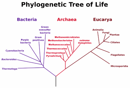
Woese and Fox’s Tree of Life, 1977

Woese’s work gave us our modern phylogenetic tree of life in 1977 (picture right). This tree showed that Archea are closer in phylogenetic history to us than bacteria. It is a good bet that both eukaryotes and prokaryotes branched off from an Archea that lived in undersea geothermal vents, making energy from hydrogen gas, warm water, and underwater nutrients richly available in those vents. Chemosynthesis, in other words, likely arrived on Earth long before photosynthesis.

What’s more, life on Earth appears to have emerged almost as soon as our planet became cool enough to support liquid water. Metal-rich Earthlike planets, with plate tectonics, plentiful water, and volcanic vents, appear to be ideal catalysts for life, and our geochemical cycles are ideal buffers and cradles for stabilizing life once it emerges. The complex set of homoeostatic protections for life on Earth, aka the Gaia hypothesis, when stated without the woo of “planetary intelligence”, appear far more developmental, from a universal perspective, than the hypothesis’s many detractors like to admit.

Woese’s work also lends credence to Alexander Rich and Walter Gilbert’s RNA world hypothesis, the idea that self-replicating RNA emerged first, before DNA and proteins. RNA is one of those rare complex chemicals that can store memory of its past evolutionary variation and self-catalyze its own replication. In other words, it is autopoetic (capable of self-maintenance and self-improvement).

Another high point is the book’s discussion of horizontal gene transfer. Amazingly, it appears that about 8% of human DNA arrived sideways in our genome, not via sex or mutation but via viral infection. As Harald Brüssow reminds us in “The not so universal tree of life,” we have not yet incorporated viruses into our current trees of life. That is a major oversight. Retroviral insertion sequences are found everywhere in eukaryotic DNA. Viruses and cells are constantly exchanging genetic material, in all species. [Brüssow H. (2009). The not so universal tree of life or the place of viruses in the living world. Phil trans. Royal Soc. of London. doi:10.1098/rstb.2009.0036]

Tree_Of_Life_(with_horizontal_gene_transfer)

Tree of life showing vertical and (a few) horizontal gene transfers. Source: Wikipedia

Our Real Tree of Life, once we draw it to include viruses, will look even more like a network than in the figure at right. The tree drawn at right is a good step beyond Woese’s 1977 tree, but it is still much too conservative. It includes no lines between eukaryotes, for example. It ignores retroviruses and other mechanisms.  See the Wikipedia article on HGT for the great variety of DNA transfer mechanisms we’ve discovered so far.

DNA is arguably still the dominant autopoetic system on our planet today. DNA’s astonishing ability to copy, vary, and improve itself, to jump around inside the cell as transposons, to jump between cells and organisms via viral and retroviral insertion, and to use vertical methods like germline mutation and sexual recombination, has made all living species on Earth much more of a single interdependent network than most of us realize.

This is an important idea to understand, because is the genetic network, not any collection of species, that has always been the true survivor and improver in life’s story. Many past environmental catastrophes, like the Permian extinction, and the K-T meteorite impact, have wiped out the vast majority of species, but I would personally bet almost all of the diversity of the genetic network survived each of those events. This is obviously true in developmental genes, which are highly conserved. If any complex species survives a catastrophe, the developmental core of all complex species survives. But I suspect it is true for most evolutionary (nonconserved) genes as well. We shall see if the evidence from modern catastrophes bears this assertion out. Genes are typically reassorted into hardier species after each catastrophe, and those species, having no competition and ample resources, make great leaps in innovation immediately after each major catastrophe. I call that the catalytic catastrophe hypothesis, and I look forward to seeing it proven in coming years.

Interdependent networks, in other words, always win out in complex selective environments, over time. Such networks are stabler, safer, more ethical, and more capable than isolated individuals. There are deep lessons in complexity science and network science to be discovered here, lessons that tell us why our leading forms of artificial intelligence later this century will be driven to not only be deeply biologically-inspired, but also ethical, empathic, and self-regulating collectives, just like us. Complex selection and developmental optima will ensure this is so, statistically speaking, in my view.

Again, if Quammen had covered convergent molecular phyogenetics, and a bit of evo-devo and developmental genetics, he would he would have given us a better set of trees and networks to ponder. If he’d wrestled with the convergent features of biological development at the organismic scale, he might have begun to recognize it at the ecosystem scale, and help us to begin to see and ponder it too.

Life is a complex, interdependent network, but it is also going somewhere. It is developing, not just evolving. I speculate on the intrinsic goals of evo-devo systems in my 2019 paper above. It may be too early to for us to say with certainty what goals life has, as a complex evo-devo network, but it is not to early to recognize that such goals must exist, both from evolutionary and developmental perspectives.

When considered as a single interdependent network, life’s story on Earth so far has been a curiously smooth and continually accelerating trajectory of increasing complexity, stability, ability, and intelligence. Something very curious is going on in all the Earthlike, high-complexity environments in the universe. We need to start recognizing and studying it much more closely if we wish to understand accelerating change, complexity and adaptation from a universal perspective, not just our own.

Obama’s BRAIN Initiative – A Poor Start On a Brain Mapping Vision

This post goes in my deviants category, as it is about someone who I believe has made an important but correctable mistake, who could know better, and who therefore deserves to be called out and reproved, so they might act better in the future.

obamabraininitiativeObama’s BRAIN (Brain Research through Advancing Innovative Neurotechnologies) Initiative, announced today, concerns what is arguably the most important scientific project we humans are doing today: figuring out how higher biological intelligence works, by exploring and mapping it in living and preserved brains all relevant resolutions. Neuroscientists have developed powerful new mapping tools and software in two main categories. Functional connectomics (also called Brain Activity Maps) is the process of mapping synaptic connectivity and neural activity to biological function, including memory, in living brains. To make these maps we have new tools for monitoring neural action in vivo at molecular, cellular and circuit levels, like optogenetics, calcium imaging, nanoparticle sensors, and other clever advances. Structural connectomics (also called just Connectomics Maps) is the process of mapping synaptic, cellular, and nuclear (epigenetic) information in chemically preserved, nonliving brains (worms, flies, snails, zebrafish, mice, monkeys, humans, etc.), as a path to figuring out function. There are also new tools and software for the automated slicing, scanning, and mapping of synaptic connections. It was observation of the rapid advances in these areas that led led Ken Hayworth and I to co-found the Brain Preservation Foundation in 2010.

The new idea is that combining these two forms of brain mapping may finally allow us to uncover the neural coding system, the ways networks of neurons store short and long term information in their association patterns and strengths. The paper that launched the Brain Activity Map proposal is The Brain Activity Map Project and the Challenge of Functional Connectomics, Alvisatros et.al., Neuron 74, June 21, 2012 (5 pp). It’s a great intro to the exciting promise of this field, and a call to action. Wikipedia has no page yet on functional connectomics (perhaps a neuroscientist will start one) but they do have a page now on the BRAIN Initiative.

There are many potential benefits to functional and structural connectomics for science and medicine, but their greatest promise, in my opinion, is that they will accelerate our ability to build intelligence in our much faster and eventually far more capable electronic systems. Some of the brain’s circuit structure and function will turn out to be highly similar from brain to brain (developmental) and some will be unpredictably different (sometimes called “evolutionary” or “Darwinian” differences). Understanding the developmental parts of the brain, and how they constrain and enable the evolutionary parts, will get us much farther down the road of building self-improving artificial intelligences. Activity and connectomics maps, and a few other new tools for monitoring neural activity at molecular scale will of course provide many medical and neuroscientific benefits, and these can be sold most easily to the general public, but the intelligence benefit for science and society, via advances in computational neuroscience and machine learning may quickly become the most important for us.

brainchangesitselfObama hinted in his State of the Union address in February that he wanted to see America’s brain-mapping and related neuroscience efforts  “reach level of research and development not seen since the height of the Space Race.”  Science writer John Markoff, in a great NYT article Feb 17th, summarized the views of the founding scientists behind the Brain Activity Map proposal, that funding on the order of $3B, or $300M/year, should be publicly committed to this project. That would make it less than the $3.8B we spent on the Human Genome Project from 1998-2003, an investment which returned, according to a 2011 Battelle report, $796B in new economic activity between 1998 and 2010. A return on investment of greater than 200, one of those rare ROIs you see when opening up an entirely new field.

Functional and Structural Connectomics promises to have that same kind of fundamental impact, opening up neuroscience and bringing all the benefits of understanding natural intelligent systems to the technology world. In addition, understanding how the brain uses connectionist features like redundancy and neuroplasticity to protect its critical functions would be huge advances for medical science and therapy. I recommend reading Norman Doidge, in The Brain that Changes Itself, 2007,  for fantastic and motivating examples of how resilient our brains can be to memory loss and damage.

Unfortunately, in his announcement today President Obama has committed just $100M to the project for its first year budget. And the money committed so far is a hodge-podge that is not project or map focused. Consider that Europe’s Human Brain Project just got $1.3B committed from the EU for the next ten years, even though that project is doing far more theoretical, lower-resolution simulation work that will be highly likely to have a much poorer payoff, in a world where we haven’t yet cracked the static and dynamic neural coding algorithms. Yes, the Human Genome Project started with the same small seed funding of around $100M the first year. But that was when genomics was untested, proteomics a dream, and understanding and mapping the brain still largely unreachable. We’re way beyond those early days now. We know how important maps are, and that we have tools available to make them, and the data sciences folks and hardware to analyze all the new public domain data that will result. It’s time to match real funds with the rhetoric.

As I said, the scientists involved in the BRAIN initiative know we’ll need at least $3B to make major discoveries with activity maps alone, and this doesn’t even include connectomics maps, which deserve a few billion as well, if we really want to figure out the neural coding language in any complex animal (say, a fly, or perhaps an Etruscan shrew, a mammal with only 1 million neurons). $5B is not a lot of money for the incredible intellectual advances we can expect. To put this in perspective, we are presently spending $85 billion per month on QE3. Obama cobbled this $100M together by redirecting existing funds in NIH, DARPA, and NSF budgets, so it isn’t even new money, it’s just reclassified R&D. An NIH working group has been designated to develop a multi-year plan with cost estimates by June 2014, and Obama has fast tracked the group by asking for an interim report by fall 2013. But its still quite unclear what the goals of the project are, and whether connectomics maps will even play a role.  If they pass on funding synapse-level connectomics maps, that will be a major failure of nerve.

Isn’t $100M a great start for Year 1? Not in my book. What would have been commendable, for a project with this magnitude of potential benefit, would have been starting with a level of finding that is ten times more, or at least a billion dollars up front, and a commitment to seek at least a billion a year for the next ten years. That’s enough to influence students to enter into this field, and would place this project in the light it deserves – one of the best science projects we could work on at this unique point in human history. We should and can demand a lot more from this second term president, particularly one who understands science and tech the way he does. Obama has committed to a commission to study the bioethical issues that might emerge (a concession to conservatives perhaps), but so far his “dream team” of 15 neuroscientists have not committed to connectomics maps, as far as I’ve read. Perhaps they will, but given the vagueness of today’s announcement, it’s quite possible we we’ll see something better in the future. But this isn’t the kind of start that inspires confidence.

Ultimately, as readers of this blog know, whether second-term American politicians have the courage to say it publicly or not yet, smarter machines, even more than adding more 20th century-style jobs, have become the primary wealth creator in the developed world, so that’s where our thoughts should go first, as we look for ways to improve our lot. I think it’s time we got serious as a species about realizing what kind of progress the universe has engaged us in. We are here to use our wits and works to become something greater than ourselves. Our highest role appears to be to take what the universe has done with us and make something even smarter, more ethical, more productive, and more resilient as our progeny. This is what civilization has been about, since the birth of technology, as I see it.

Want to let the Obama administration know your thoughts on making Brain Mapping, including connectomics maps, a top funding priority? You can send a brief email to the White House by using this form, as I have. Thanks.

A "clarified" brain (lipids removed, everything else in place). Transparent to optical microscopy, all the proteins, receptors, RNAs able to be repetitively interrogated with molecular probes. Amazing!

A “clarified” mouse brain at right (lipids removed, all else stays in place). Transparent to optical microscopy, all proteins, receptors, RNAs can be repetitively interrogated with molecular probes. Amazing!

4/18/2013 Update: The Stanford press release on 4/10 announcing CLARITY, the Karl Deisseroth lab’s amazing new method for optically transparent brain mapping, just makes what I said above more appropriate and urgent, from my perspective. Deisseroth is one of the 15 experts on Obama’s neuroscience dream team, so I’m sure he advised the White House of its implications. The CLARITY paper was accepted for review at Nature in September 2012. The CLARITY method is like PCR, a multipurpose, revolutionary new research tool that will open up vast new imaging and molecular phenotyping research capabilities in any biological tissue, and in particular the brain. Salk’s Terry Sejnowski said: “It’s exactly the technique everyone’s been waiting for.” He told the Associate Press that it will speed up brain anatomy research by “10 to 100 times.”

And yet Obama’s team still proposed just $100M in funding for brain mapping for the first year. That’s simply ridiculous. Please, America, wake up! It’s time to spend some real money on neuroscience and bust humanity out of its ignorance. Stop being scared of how much better things will soon be, once we’ve cracked the riddles of neural information processing. Someone also needs to give Deisseroth a serious prize or two. Optogenetics and CLARITY, both out of his lab, are each profoundly important biological sciences breakthroughs.

10/27/2013 Update: Ugh! Obama’s BRAIN initiative (April 2013) has barely started and it’s already been co-opted. Politics is not pretty. http://www.nytimes.com/2013/10/25/science/pentagon-agency-to-spend-70-million-on-brain-research.html?_r=0

DARPA will spend their chunk of the funds (half of the money we’ve committed so far to the initiative, $70M over five years, which is peanuts, as I’ve said before) on a very-low-yield clinical project (deep brain monitoring and stimulation) vs a multipronged effort to improve human brain structural and functional connectomics (circuit tracing, electrical activity mapping, optogenetics, nanosensors). The potential for brain mapping as the #1 focus of the initiative is gone, mere months after they announced it.

Apparently Obama got the wrong partners (DARPA, NSF, NIH) together for his BRAIN initiative. The 2012 Brain Activity Map proposal that Alvisatros, Church and others made to the White House was all about functional connectomics. This has now taken a back seat to deep brain stimulation and monitoring experiments. Drats! I like DARPA, but I’m sure that initiative mostly won’t work, without functional maps, and I’m not even a neuroscientist. But DARPA likes clinical work with near-term potential benefit (or at least the potential promise of it). It would have taken a firm hand to keep them focused on Brain Mapping, which is the real prize accessible to science, at this stage of our collective technical abilities (more accurately, ineptitude) when it comes to the brain. That leadership is missing today.

We need a lot more money, at least a billion dollars a year, devoted to funding the Basic Science of structural and functional Brain Maps, not these expensive clinical junkets. How else can we solve the memory code, and thereby understand how neural nets actually work, and thus make better AIs? Or, as my friend Steve Coles, MD, PhD says, what is the genetic reason why we humans have a Broca’s area and chimps don’t? What is the connectomics of higher intelligence? All most future-important questions about human, social, and machine intelligence are dependent on better brain maps. Ken Hayworth and I started the Brain Preservation Foundation (http://www.brainpreservation.org) in 2010 with the realization that these maps are coming, and will greatly improve our understanding of who we are, and what we can do with our memories and identities after biological death.

One fine day we can expect a real Human Brain Mapping initiative, one that really does greatly improve our collective understanding of the brain, for all humanity, for all time. Just like the Human Genome Project uncovered the epigenome and illuminated the proteome, and now we need Human Epigenome and Proteome Projects, which also haven’t materialized, because we are so broke and unmotivated to do Big Life Science.

The world needs Brain Maps, Epigenome Maps and Proteome Maps as the new science moonshots for the next five to ten years. These would be completed under budget and under time with more powerful computers than we expected, just like the HGP was.

In the meantime, we get this BRAIN initiative elephant, designed by committee. This is a major loss of vision and leadership here. #ObamaFail

Vote for scientific and technical leadership in 2016, irrespective of party. It’s high time we get some representatives who see, sooner or later, how extraordinary humanity’s future will be. Sooner would be nice, eh?

Preserving the Self for Later Emulation: What Brain Features Do We Need?

Let me propose to you four surprising statements about the future:

1. As I argue in this 2010 video, both chemical and cryogenic brain preservation technologies may soon be validated to inexpensively preserve the key features of our memories and identity at our biological death.

2. If either or both forms of brain preservation can be validated to reliably store retrievable and useful individual mental information, these medical procedures should be made available in all ethical societies as an option, for those who might want it, at biological death.

3. If neuroscience, biologically-inspired computer science, microscopy, scanning, and robotics keep improving as they have so far, preserved human memories and identity may be affordably reanimated by being “uploaded” into computer simulations, even before the end of this century. Such “uploading” is being attempted for specific memories in animals today, and new tools like optogenetics and expansion microscopy cause leading neuroscientists to expect they will soon decipher and be able to manipulate (removing and introducing new memories) the neural code.

4. In all societies where a significant minority (let’s say 100,000 people) have done brain preservation at biological death, significant positive social change may result in those societies today, regardless of how much information is eventually recovered from preserved brains.

These are all extraordinary claims, each requiring strong evidence. Many questions must be answered before we can believe any of them. Yet I provisionally believe all four of these statements, and that is why I co-founded the Brain Preservation Foundation in 2010 with the neuroscientist Ken Hayworth. BPF is a 501c3 noprofit, chartered to put the emerging science of brain preservation under the microscope. Check us out, and join our newsletter if you’d like to stay updated on our efforts.

[2018 Update: For the latest on the neural code, the physical storage of memories in the brain, see this excellent paper by Carillo-Reid et al. (2018). It show how we are now imaging and optically manipulating neural ensembles, small spatiotemporally connected networks of neurons that link to other ensembles and are the building blocks of neural circuits. Neuroscientists believe these ensembles and circuits store engrams (memories and models) in the brain. With new optogenetics tools, we can find and manipulate these ensembles, and possibly create new ones, which could then be recalled by selectively stimulating any individual cell in the ensemble. This would be a true demonstration of memory creation, and major advance in deciphering the neural code.

More hints that neural synchrony in the claustrum is the root source of consciousness came in 2014, when a neurologist at GW School of Medicine, Mohamad Koubeissi, managed to turn off and on consciousness in a single patient by electrically stimulating her claustrum with deep brain electrodes. Electrical stimulation of brains has a long history in neurology and neuroscience, but it had never resulted in consciousness control, until now. Then in 2017, an fMRI study in awake and anesthetized rodents showed strong interhemispheric functional connectivity, via the claustrum, to both the prefrontal cortex and the thalamus in the awake state, dynamic connectivity that was lost in the anesthetic state. This was the first study to identify specific neural substrates by which the claustrum may “orchestrate” (to use Francis Crick’s term) consciousness. This work is a major vindication of Crick and Koch’s 2004 hypothesis that the claustrum is the central creator of consciousness. We are truly on the edge of understanding it as a fully mechanistic process. Understanding consciousness mechanistically will allow us to ask how we might go about instantiating it into nonbiological intelligence systems, perhaps even some time this century.

There are also some speculative hints, in 2018, of a possible epigenetic “backup” storage of memory. Some very old experiments by James McConnell in 1959, recently redone by Michael Levin at Tufts and David Glanzman at UCLA, tell us our memories may not only exist in our synapses and dendrites, they may also be stored, to some degree, epigenetically in our neural nuclei, via modification to the histone proteins that wrap DNA and regulate its folding. Having such a digital memory backup system would be clever, if brains and evolution figured out how to do that. A readable “backup” could could be used to reestablish the right connections if they are disrupted, and would our memories particularly robust to biophysical stresses that deform the analog morphology of neural networks in a living, moving brain. If some degree of epigenetic memory backup (to neural nets) exists and is extensive, it might mean a lot of a person’s memory could be retrieved and uploaded in the future, even if even if a person was preserved using poor methods (like most cryonics patients today), or preserved many hours after death (as their neural morphology starts to decompose). More research is going to be needed to determine if such a system exists, and if so, how many hours it might survive after death.]

For an excellent primer on the latest neuroscience theories and computer models in how the brain works, read the free wiki book, Computational Cognitive Neuroscience, O’Reilly and Munakata (2012). Chapter 8, Learning and Memory, is excellent. To understand how and why “you” might survive being uploaded into a computer or robotic body, if that was easiest way to bring you back to the world in the future, see BPF Advisor Mark Walker’s article, “Personal Identity and Mind Uploading,” JET, 22:37-51, 2012.

In this post, I’d like to give you my best provisional answer to a question relevant to the first three statements above:

To preserve the self for later emulation in a computer simulation, what brain features do we need?

We can distinguish three distinct information processing layers in the brain:[1]

1. Electrical Activity (“Sensation, Thought, and Consciousness”)
These brain features are stored from milliseconds to seconds, in electrical circuits.

2. Short-term Chemical Activity (Short- & Intermediate-term Learning – “Synapse I”)
These brain features are stored from seconds to a few days in our neural synapses (synaptome), by temporary molecular changes made to preexisting neural signaling proteins and synapses, as well as temporary chnages to the neural epigenome (DNA transcription and inhibition machinery, an overlay on the human genome).

3. Long-term Molecular Changes (Long-term Learning – “Nucleus and Synapse II”)
These are stored from years to a lifetime in our neuron’s connectome, synaptome, and nucleus (epigenome), by permanent molecular changes to neural DNA, the synthesis of new neural proteins and receptors in existing synapses, and the creation of new synapses.

All three of these brain processes involve a still-imperfectly understood combination of both digital (on or off, discrete states, including epigenetic DNA states and neural firing states) and analog (continuous, spatially related, wavelike) information and computational processes, which integrate in a statistical, associational, competitive, and massively parallel manner. At present, it is a reasonable assumption that only the third layer listed above, where long-term durable molecular changes occur, must be preserved for later memory and identity reanimation.

The following overview of each of these three key information processing layers should help explain this assumption.

1. Electrical Activity (“Sensation, Thought, and Consciousness”)

Our electrical brain includes short-distance ionic diffusion in and between neurons and their supporting cells (i.e., calcium wave communication in astrocytes), action potentials (how neurons send signals from their dendrites to their synapses), synaptic potentials (how signals cross the gaps between neurons), circuits (loops and networks) and synchrony (neurons that fire in unison, though they are widely separated). Electrical features operate at very fast timescales, from milliseconds to a few seconds, and are variable (not exact), volatile, and easily disrupted.

Neural Synchrony – Our Leading Model of Higher Perception and Consciousness . Image: Senkowski et.al., 2008

These features certainly feel very important to us. They include our sensations (sensory memory) and current thoughts (commonly called “short-term” memory by neuroscientists). Recurrent loops, special electrical circuits that cycle back on themselves, hold our current thoughts (when you rehearse some information to avoid forgetting it, you are literally keeping it “in the loop”). Neural synchrony is my favorite current theory (among several currently in competition) for the medium that creates our conscious perceptions. When it happens in the self-modeling areas of our brain, it gives us self-aware consciousness.

Yet electrical features are also fleeting. When you sleep, or are knocked unconscious, or are given an anesthetic, your consciousness disappears, only to be “rebooted” later, from more stable parts of your brain. Our memories aren’t even recalled with precision but are rather recreated, as volatile electrical processes, from these molecular long-term stores, in ways easily influenced by our mental state and cognitive priming (what else is on our mind). That’s why eyewitness testimony is so variable and unreliable.

The electrical features of our self are thus like the “foam” on the top of the wave of our long-term memories and personality. They make us unique for a moment, as they hold only our most immediate thinking processes.[2] Amazingly, people who undergo special surgeries that stop their heart, and some who drown in very cold water, can have no detectable EEG (electrical patterns) for more than thirty minutes, and their brains successfully reboot after rewarming them. Essentially, these individuals are recovering from clinical brain death. Not only do they not have consciousness during this period, they have no unconscious thoughts. Yet because their deeper layers aren’t too disrupted, they can restart their electrical activities.

An excellent though very technical book about neural spikes, loops, and synchrony is Rhythms of the BrainGyorgy Buzsaki, 2006. It explains the emergent properties and integrative functions of these “highest order” electrical features of our brain. See also this recent discovery of electric field coupling among neighboring neurons, by leading neuroscientists Henry Markram, Christof Koch, and others, and reported by Peter Hankins on his great cognitive science blog, Conscious Entities. There are some big mysteries still left to uncover regarding synchrony. Ephaptic coupling is a way for neurons to synchronize spike timing in neighboring neurons, via a mechanism completely independent of synapses. Neurons are much more versatile in both modes of communication and synchrony than previously thought.

My late mentor at UCSD, Francis Crick, and his brilliant Caltech collaborator, Christof Koch, call this topic the search for the Neural Correlates of Consciousness. It’s a great phrase. Consciousness is not a mystery we’ll never solve, but according to a number of neuroscientists it is a physical process of neural synchrony, in particular regions of your brain. The region Crick and Koch was most interested in was the claustrum, a thin sheet just above the putamen, near the center of the brain, found in all mammals, which  has many longitudinal tracts that connect to many areas of the cerebral cortex and to the thalamus, the central relay station in the brain. In 2005, the year after Crick’s death, Koch published their hypothesis that the claustrum is the central synchronizer of conscious experience.

These brief, rhythmic synchronizations share information between highly different, specialized groups of neurons in distant regions of the brain by tightening up (“binding”) their interdependent sequences of action potentials. The synchronizations are controlled by the inhibitory neurons in our brain, which use the GABA neurotransmitter. Disrupt gamma synch, as with anesthesia, and you take away consciousness. Give a drug like zolpidem, which activates GABA neurons and increases gamma synch, to patients who are in persistent vegetative state, and amazingly, you will wake 60% of them up from their comas, to varying degrees!

Wikipedia doesn’t yet have a good explanation of the gamma synchrony model of consciousness, but they do have a good page on the claustrum. Laura Colgin at Kavli has found two reliable gamma synch mechanisms in rat hippocampus. She speculates that slow gamma makes stored memories available to current consciousness, and fast gamma integrates sensations to create conscious perceptions. 

In sum, though neuroscientists don’t yet all agree on many of the details, many have found neural correlates of sensations, thoughts, emotions, and consciousness in the electrical features of our brains. In conjunction with the short-term chemical changes we will describe next, these processes represent both our “highest” (most valued) and yet at the same time, our most volatile and least-unique self.

It is quite likely, in my view, that if we uploaded your brain into a computer, and then reestablished different consciousness network states than the ones that existed in your biology, at death, states that were an “average” of typical human networks, you would still wake up feeling like essentially the same “you”, and others would describe you as the same as well. Consider that we all have different kinds of consciousness on a daily basis.

In other words, the dynamic specifics of our consciousness networks (which we don’t yet deeply understand), as opposed to their structural connectivity with the rest of the brain, (which we are now identifying in areas like the claustrum, and know how to preserve) may be the least important contributors to our unique identity.

While for many of us, consciousness is the feature of our minds that we love the most, it appears its primary role is to be an “orchestra conductor” of much more carefully stored, and slower-changing “lower-level” layers of you. If you change orchestra conductors, you will still get a symphony that is beautiful, and largely the same.

Let’s look at those significantly more unique lower layers now.

2. Short-term Chemical Activity (Short- and Intermediate-term Learning – “Synapse I”)

Short-term chemical activity is the next layer down. It involves all our short- and intermediate term learning and memory, everything beyond our sensations, current thoughts, and consciousness, but not including our long-term memories. We can call this layer “Synapse I.”

As our electrical experiences and thoughts race around the various circuits in our heads, we make a number of short-term learning changes in our neural networks to capture, for the moment, what we’ve just experienced and learned. These involve changes to preexisting proteins in our preexisting synapses (communication junctions), changes that last for minutes (short-term) to days (intermediate-term). These are changes in both the mechanics of neurotransmitter release and short-term facilitation (strengthening) or depression (weakening) of synaptic effectiveness. Synapses are temporarily modified by the precise timing and frequency of electrical signals (action potentials) received by the postsynaptic neuron, a process called spike-timing dependent plasticity. There are short-term changes in signaling molecules (neurotransmitters, cAMP, Ca++, CamKII, PKA, MAPK), and membrane receptors (NMDA). Phosphorylation states (chemical tags) are altered on some of these molecules, and a temporary equilibrium between kinases (enzymes that add phosphates to key molecules) and phosphatases (enzymes that take them away) is established in the synapse.

[Note: In late 2012, Ye et. al. showed in Aplysia how precise spatiotemporal signaling in the synapse involving PKA holds short-term memories in synaptic electrochemical networks, and the interaction of PKA and MAPK holds intermediate-term memories in these networks, in a process called synaptic facilitation.]

Throughout our day, and particularly when we sleep, our short- and intermediate-term brain writes important parts of its experiences to our long-term memory (the subject of our next section), building durable new synaptic connections, so this learning can stay with us for years to life, in a process called memory consolidation. Memory consolidation seems to happen best when we are in slow wave (deep and dreamless) sleep, which we get in cycles during the night (and especially well if our sleeping room is dark and quiet) and also during a good nap. That’s why a short nap after an intense learning session is considered a great way to “lock in” what you’ve learned.

When any of our short- or intermediate-term memories or thinking patterns are selected to be written into long-term memory, communication with the cell nucleus must occur, and new membrane proteins and synapses are then built, involving new or altered circuits in the connectome. If not, the new memory or pattern dies out.[3]

Again, long-term memory formation moves a subset of our recent learning and memories, apparently the most relevant parts, from temporary spatiotemporal signaling states to permanent new synaptic structures, anchored to the cytoskeleton of each neuron. We can think of the new proteins, synapses, and circuits established in long-term memory in neural synapses and nuclei in a way that is very roughly like DNA, as they are long-term stable structures, encoded in a partly digital, partly analog form, that will endure all the flux and variability of the biochemistry within each neuron, over a lifetime.  It is these unique long-term synaptic and epigenetic networks that we must preserve, scan, and upload in creating neural emulations, as we will discuss.

Now consider this key insight about short- and intermediate term (Synapse I) learning, which helps you appreciate it is not consciousness, and it is not your long-term memory, but something altogether different. Subjectively, we all know we can store both a far greater total number and a far greater temporal density of episodic (experiential) and declarative (factual) memories in our short-term memory, than we can in long-term memory. That’s what we do when we cram for a test, intensively read important information, engage in intense discussions, or pay for great experiences, as we have learned we can greatly alter our short term memory, and we know having a lot of relevant, motivating information “loaded” into our short term memory can be a great help to us as we work on managing both our long-term strategies and our daily tasks.

Most of this short-term information is not selected to be written into long-term memory, as far as both our subjective experience and neuroscience research knows today. It has a half-life of days, and it steadily decays after it is learned. But all that memory can still be highly useful in the moment. It plays a key role in regulating what we can think and do, on any particular day.

Given what we’ve seen so far, do you think our short-term (Synapse I) learning needs to be preserved for you to return to life as essentially the same person you were when you died? I don’t think so. Claiming it does seems to me to be an extreme claim.

Consider that if your NMDA receptor distributions aren’t recoverable from a future emulation, for example, you should only have lost the last couple days of your life experience prior to being preserved, in our present understanding of how these systems are most likely to have evolved and operate.

I would expect any good reanimation program could use any baseline (species average) version of such receptors and you’d wake up, as almost entirely the same “you”. Perhaps we’ll need another decade or two of neuroscience to definitively answer such questions, but we can already have good reasonable intuitions about them today.

Let’s look a bit closer at how neurons work to understand the amazing capacity of short-term memory in a bit more detail.

Neural dendrites, cell body,  action potential, and synapses. Image: Gallant’s Biology.

All our neurons work in circuits, and strengthen or weaken their connections based on chemical and electrical activity, in a process called Hebbian learning. Just like your muscles, which come in two sets that oppose each other around every joint, neural circuits are both excitatory and inhibitory at many decision points in the network. One of the most important decision points is the cell bodies of each neuron, where the nucleus is. The electrochemical current from all the dendrites (“roots”) of each neuron flows toward its cell body, and action potentials (current waves) flow from the cell body to its synapses (“branches”), along the axon (“trunk”) of each neuron. Glutamate is the main neurotransmitter we use to send excitatory current from a synapse to the dendrite of the next neuron in a circuit (the postsynaptic neuron). Glutaminergic synapses are thus called “positive” in sign, and they promote electrical activity throughout the brain. GABA is the main neurotransmitter we use to let inhibitory current leak out of a postsynaptic dendrite. GABAergic synapses are thus called “negative” in sign, and they depress circuits throughout the brain. With few exceptions, neurons use just one type of neurotransmitter, or the same small set of neurotransmitters, at all their synapses.

Electrically, each neuron sums the net result of the positive and negative inputs it receives from all its dendrites, over milliseconds to seconds. As part of this summing process dendrites also make their own local and weak types of mini-action potentials, dendritic spikes, and we’ve recently learned they use these mini-spikes to do complex information processing prior to sending current to the cell body. This reminds us that computationalists still have a good ways to go before they can build neural network models sufficiently complex to honor reality. Then, if the current received at the cell body exceeds that neuron’s threshold,  it sends a traditional action potential (depolarizing electrochemical signal, or “spike”) to all its synapses. As the brain learns, our synapses enlarge or shrink, giving them greater or lesser excitatory or inhibitory effect, and we will either grow more or lose our synapses, depending on the value of the circuit.

The architecture of memory, thought, emotion, and consciousness may thus be reducible to a surprisingly simple set of algorithms, connections, weights, signaling molecules and electrical features in each neuron, working together in a massively parallel way to create computational networks that are far more complex than the individual parts.

Hippocampus and frontal lobes. Image: NIH

In higher animals, the neurons in our hippocampi (two c-shaped areas of ancient, primitive, three-layer cortex in each hemisphere of our brain), and the connections they make to the rest of our cerebral cortex (especially to our frontal cortex), store all kinds of episodic (experiential) and declarative (fact-based) information, all from our last few days of life. At the same time, neurons in our cerebellum (a more primitive, “little brain” at the base of our skull) store procedural learning and memory (how to move our bodies in space). Experiments with rats and primates tell us that each hippocampus makes perhaps tens of thousands of new neurons every day, from neural stem cells. Other than for repair after certain kinds of injury, no other part of the adult brain is able to use stem cells in detectable numbers, as far as we know. The rest of our brain is postmitotic (unable to use cell division to maintain its structure), as neuroscientists demonstrated in an elegant experiment in 2006. Our neurons must be maintained by our immune and repair systems, and as they die via natural aging, or kill themselves in apoptosis, memories start to die.

Hippocampal dendritic spines. Image: Fiala & Harris, 2000.

Our hippocampal neurons may be the primary place where we temporarily hold, in the uniquely dense synapses of this evolutionarily older cortex, and via their connections to the rest of our evolutionarily newer cortex, much of the information we have learned over the last day or two, during our entire adult life.

At right is a picture of a computer reconstruction of a small section of ten columns of synapse-rich “spiny dendrites”, from the CA1 (input) region of the hippocampus. CA1 contains areas like place cells, imprinted genetically with detailed maps of 3D space. Like the digestive cells lining our gut, and the skin cells at our fingertips, certain hippocampal neurons appear to get worn out on a regular basis by this demanding short-term memory holding function, and so some neuroscientists think new ones must regularly grow and mature to replace them.

People whose hippocampi are both surgically removed, like the memory disorder patient Henry Moliason, who had this done at the age of 27, can’t update their long-term episodic and declarative memories. H.M.’s long-term memory and personality was mostly “frozen” at 27. He could occasionally add bits of new information to long-term memories of the same type he’d built before the surgery, and he could learn new procedural (spatial and muscle) memories in his cerebellum, but he had no cerebral knowledge that he’d added these memories. H.M.’s amazing life suggests that if the brain preservation process damaged our hippocampi, but not the rest of our brain, we’d come back without our most recent experiences (two-day amnesia), but all our older memories and personality would still be intact.  Ted Berger at USC managed to build a simple version of an artificial electronic hippocampus for mice in 2005, so there’s a good reason to believe that this part of our brain, though important, isn’t irreplaceable. As long as you could install an artificial hippocampus in the computer emulation constructed from your scanned brain, you’d be back in business as a learning organism, with only some of your more recent memories and learning erased. This all helps us understand that what cognitive scientist Daniel Dennett would call our center of narrative gravity, our most unique self, is our long-term memory.

The fact that only special areas of our hippocampus can add new cells during life exposes a harsh reality about our biological brains. We are all born with a large but fixed memory capacity, both short-term and long-term, and this capacity gets increasingly used up, pruned and potentiated, the older we get. Anyone over 40, like myself, knows they are considerably less flexible at learning new things than they were at 20. That decreasing flexibility is simply a result of the physics of network formation in finite biological brains. We can still add new branches, and new connections, but it gets harder over time.

Now we arrive at our truest self, the part I will argue that we care most about preserving and sharing with our loved ones and society. It is this self that I expect will later merge with the Digital Twin (to be discussed shortly) that many of us may leave behind for our loved ones after our deaths in the 2020’s and beyond, as strange as that concept might sound today.

Experience-based learning. Image: Graham Paterson, Children’s Hospital Boston

3. Long-term Molecular Changes (Long-term Learning – “Nucleus and Synapse II”)

The production of long-term memory, personality, and identity requires all the short-term synaptic changes above, plus permanent molecular changes in the neuron’s Nucleus (DNA and its histones, or wrapping proteins), and the permanent creation of new cellular proteins, synapses, and circuits (Synapse II). Here’s a brief summary of our understanding of the process[4]:

3A. Nucleus (“Genome, Transcriptome, and Epigenome”)
1. Retrograde transport and signaling from the synapse to the nucleus
2. Activation of nuclear transcription factors and induction of gene expression
3. Chromatin alteration and epigenetic changes in gene expression (gene-protein networks)

3B. Synapse II (“Connectome and Synaptome”)
1. Synaptic capture of new gene products, local protein synthesis, and seeding of new synaptic sites
2. Permanent synaptic changes, activation of preexisting silent synapses, formation of new synapses.

We used several “-ome” words above. Let us briefly consider each. They are very roughly ordered below in terms of their likely contribution to our unique self, from least to most important:

The Genome. These are inherited genes and gene regulatory networks that control instinctual behaviors. Our genome includes the unique alleles we received from our parents. It is easy to preserve, as it is the same in all cells. With one tissue sample we can create a clone later, either physically, or far more likely, in a computer simulation. But this clone has only our inherited uniqueness. We’ll need contributions from the next four “omes” to add our life memories and learning to the emulation.

The Transcriptome. This is the set of proteins made (transcribed) by cells. While proteomics (another “ome” word) is in its infancy, scientists estimate each of our cells has the DNA to express ~20,000 basic protein types. Each type can be further modified after creation by adding or removing chemical tags like phosphate, methyl groups, ubiquitin, and other small molecules, so that more than a million protein subtypes may exist in a typical human body. Fortunately, each of our ~220 cell types only uses around 5,000 of these 20,000, and perhaps less than 2,000 of the 5,000 are unique to each cell type. Neurons and glia, the cell types we are most interested in, may use just a few hundred protein types to store our higher learning and memory in the nucleus and synapses. The other proteins are there to keep all of our cells alive, which is a critical precondition to being able to store long-term memories in a special subset of neural structures. All this suggests the proteomics of memory and identity, and of later memory and identity reconstruction from scanned brains, are not impossibly complex, but rather highly challenging, fascinating and eventually solvable problems.

The Epigenome. Our epigenome is a gene-regulatory layer that involves chemical changes, mostly methylation and acetylation, to DNA and to the histone proteins that wrap and expose DNA in the cell nucleus. These changes determine how DNA, RNA, and protein are expressed in the nucleus, and how neurons manage their synapses as they grow and learn. These are learning-based changes in gene-protein networks that occur during the life of the organism.  Epigenetic changes during biological development are responsible for the different transcription patterns that emerge as cells divide, turing them into the various cell and tissue types in our bodies. The Dutch famine of 1944 and the Överkalix study in Sweden tell us that some epigenetic changes can be inherited in humans, so we all should seek good nutrition and avoid toxin exposure, as we may pass some of that to our children in the form of compromised and undermethylated epigenomes. There is a lot more to the epigenome story still to be uncovered, as this 2011 article on epigenetic regulation in learning and memory in Drosophila makes clear.

 [2015, 2018 Updates: Dr. David Sweatt at U. Alabama, Birmingham has for the last few decades been one of the leaders in researching the epigenetics of memory. See this brief article by Sweatt (Exploring the Building Blocks of Memory, UAB’s The Mix, Oct 2015), for a recent update. Unfortunately, as a species and in our leading countries we spend a ridiculously small amount of money trying to uncover those building blocks, so progress has been far slower than it otherwise could have been. Also, read this great 2018 ScienceNews article, and see Wikipedia’s page on the epigenetics in learning and memory to track this ongoing story. Perhaps you’d like to become a researcher in this area yourself? You’d do us all a great service.

The human brain is the most complex piece of matter in the known universe. The question of how memory is stored is arguably the most interesting of all brain questions, and one whose answer will improve our civilization in countless ways. You’d think it would be among our top priorities, but unfortunately, it still isn’t. We are stumbling and dragging our feet into a better world, not running toward it. 

How much of the epigenome will we need to preserve to be able to recreate higher human memories in computers? That question isn’t yet clear. Epigenomic changes that direct permanent protein changes in the neural nucleus may very likely be a redundant form of memory storage.  I would currently bet that some (low?) level of epigenomic data, in concert with connectomes and synaptomes (discussed next), may be necessary to recreate our higher memories. We shall see, as they say.]

The Connectome. This is a map of our neural cell types, and how they connect. Our connectomes and much of our dendrite structure is very similar in all of us. This shared developmental structure makes it easy for us to communicate as collectives, for ideas or “memes” to jump from brain to brain. Yet with 100 billion neurons making an average of 1,000 connections to other neurons, and most of these not being developmentally controlled, we’ve got the ability to make 100 trillion connections, the large majority of which will be unique to each individual.

The Synaptome. These are key features of the ~1,000 synapses that each neuron makes to others. They are the particular long-term molecular features that determine the strength and type of each synapse, its signaling states and electrical properties, as we’ve described them above. The synaptome is the weight and type of the 100 trillion connections described above, and this information may be the most important “recording” of our unique self. Fortunately, because memories are stored in a highly redundant, distributed, and associative manner in our synaptic connections, our synaptome is to some degree fault tolerant to cell death. Both artificial and biological neural networks experience graceful degradation (partial recall, incremental death) of higher memories as individual neurons die. We also know the molecular code of long term memory is fault tolerant to the noise, deformations, and chaos of wet biology. The feedback loops between the electrical and gene-protein network subsystems interact somehow to stabilize long term memories in a special subset of durable molecular changes, in spite of all the other biochemistry furiously going on to keep the cell alive. [2015: For more on synaptic diversity, a topic we still have not fully characterized, see synaptome researcher Dr. Stephen Smith’s excellent presentation, The Synaptome Meets the Connectome, YouTube, 2012. Understanding synaptic diversity is likely to be a key piece of the memory encoding puzzle. We finally have the research tools and a modicum of funding to investigate it, via such paths as the BRAIN initiative, which so far has been vastly underfunded and overhyped, and via much more effective yet relatively small independently-funded groups, like the excellent Allen Institute for Brain Science.]

Lifelong Learning, and Your Digital Twin 

Given all this, if we want to be lifelong learners in a world of accelerating technological and job change, it is critical to get an early education that is as categorically complete (global, cosmopolitan, and scientific), moral (socially good, positive sum) and evidence-based as possible. Our children need the best mental scaffolds they can get early on, or they’ll spend the rest of their lives trying to prune away harmful and untrue thoughts and beliefs acquired in their youth. Psychologists have long known that it is much easier to add increasing specificity to a neural network than it is to unlearn (depress) any branch, once it’s built. We need to be careful about what we allow into our memory palaces.

That said, children also benefit greatly from freedom, early on in life, to study what they themselves desire to learn, and to have a good degree of control over learning outcomes and style. This freedom, and appropriate rewards for effort of any kind, induce them to build intricate mental specializations in areas they are personally passionate about. For those who want to know how to implement a 50/50 balance of broad, state-mandated learning in future-critical STEM fields, analytical thinking, and civics (the “hilt of the sword”, technical ability and broad world knowledge), and a personalized program of student-directed specialized learning, creativity, and play in the other half of the time, mastering whatever they can convince their teachers is worth studying (the “blade of the sword”, passionate specialization), I strongly recommend The Finland Phenomenon, 2010 . This exceptional film (free on YouTube now too), along with Pasi Sahlberg’s Finnish Lessons: What Can the World Learn from Educational Change in Finland, 2011, and Tony Wagner’s Creating Innovators, 2012, all demonstrate key elements of the future of learning in enlightened societies, in my opinion. It may take 20 years for the evidence to be incontrovertible, but you can give it to your child now, if you find it appealing. The US will eventually realize that if the Finns did it, rejuvenating their previously failing education system over a twenty year period, we can too.

Digital Twins – Virtual Assistants (Smart Agents) With Simple Models of Our Interests, Will Be Useful for Many of Us By the Early 2020’s. Image: MyCyberTwin.com

It is also liberating to realize that while our biological brains are less able to learn fundamentally new things as they age, all the digital technologies we use, technologies which will bring our emulations back at an affordable price later this century, will continue to get exponentially more powerful every year. Most of us don’t realize this, but everyone who uses a social network, email, or any other technology to capture things they say, see, and write about is also creating a digital simulation of themselves.

Some time in the 2020s, I expect that many of us will be talking to and with our best search engines in complex sentences (the conversational interface), and will be using personalized smart agents, “Digital Twins” (hereafter, “Twins”), which will have crude maps of our interests and personality, so they can serve us better.

[2018 Note: I now call Digital Twins by a more generic name, Personal AIs (PAIs). see my Medium series on Personal AIs for more on this amazing, accelerating, and still-little-discussed aspect of our personal and social futures.

Computational linguists know that if you capture what a person says for just two years, we are so repetitive about what we care about that a Twin could whisper into our ear the word that natural language processing algorithms predict we want when we are having a senior moment, and they’ll be right most of the time. That’s how repetitive we are, and how good natural language understanding will be by 2020. As I wrote in 2005, people who don’t run Twins will be much less productive, so they’ll be very popular, even though they’ll bring lots of new social problems in their first generation.

Now here’s a kicker: These simulations likely won’t be turned off by our loved ones when we die. It will cost little to keep them running all the time, watching what we’re doing, and compiling at-first-primitive “suggestions” for us, and our children, friends, and colleagues will occasionally use them to interact conversationally, and only in appropriate contexts, with these semantic simulations, to keep the best of our thoughts, experiences and personalities accessible to them when desired. Of course, many folks will be creeped out by this idea, but others will find it a way to reduce the great pain of losing our most loved individuals to biological death.

Once folks realize that their Twins really are a kind of “digital immortalization” of parts of themselves, and once neuroscience has proven that we can read (“upload”) simple memories from preserved and scanned animal brains, at that point preserving one’s brain for later uploading into a Twin may seem an increasingly obvious and responsible choice for dying individuals, especially if the cost to do so is quite affordable. It will eventually be covered by health care in our wealthiest societies.

What’s more, recent advances in molecular scale MRI scanning strongly suggest that future scanning technologies should be able to nondestructively scan entire preserved brains, to upload their molecular states, memories, and higher functions. So if the first scan isn’t perfect, it can always be updated later, from the preserved physical brain.

We can see that teaching our children and ourselves to be digital natives and digital activists, to use the social web and the first affordable commercial lifelogs when they arrive, is one important way for us to build an ever more capable Twin for ourselves and for our loved ones (after we die), even as our biological self naturally slows down and simplifies (prunes away branches of knowledge and memories we once had ready access to) with advancing age.

To Understand Intelligence and Learning – Start with Single-Celled Organisms

Single-celled animal. Image: Anthony Horth

To understand how these subsystems interact in a living organism, let’s start in as simple a model organism as we can find, single-celled animals, organisms that don’t even have nervous systems as we know them. Wetware, Dennis Bray, 2009 is a great tour of these animals. Single-celled eukaryotes like Stentor, Paramecium, and Amoeba do complex information processing, and hold short-term memories in their chemical networks. In 2008, we learned that Amoeba remember and anticipate cold shocks, for example. These networks include the cell’s genome, epigenome, cellular proteins, cytoskeleton, receptors, and cell membrane. They are true computational networks, with both neural-network like and Boolean logic properties. Genes and proteins integrate signals from other genes and proteins, and selectively switch and transmit signals, just like neurons do. The genes in each cell, via RNA, determine which proteins are made, when and where. Most protein changes are part of the short term computation being done in a cell, but a special few will lead to lasting changes in the epigenome and the cytoskeleton and receptors in and on the surface of the cell. These long-term changes are the ones we care most about, as they store the cell’s unique memory and identity.

Until computational neuroscience[5] can predictively model how the gene-protein networks in a Paramecium allows these animals to evaluate options, assign priorities, regulate their moment-by-moment computational attention, continually vary strategies for chasing prey and avoiding toxins, and chemically store their representations, habituations, and memories in an intracellular environment, all within a single cell that has no proper nervous system, the field will be missing its Rosetta Stone. Electrical waves exist in these single-celled animals, but with the exception of mitochondrial energy production, they are of the most primitive, diffusion-based kind. All the considerable intelligence in these animals is coursing, moment by moment, through their gene-protein networks.

BPF Advisor Randal Koene likes to use the phrase “Substrate-Independent Minds” to talk about uploading. One big step to realizing how achievable uploading will likely be involves understanding the patternism hypothesis, and recognizing some of the ways nature has already built substrate-variant “minds” in complex organisms that arrived before brains. We’ve just discussed surprisingly smart neuron-independent “minds” in some single-celled organisms. Many species of plant also have very complex “thinking” abilities, all without the use of neurons.

Take a look at the Plant Intelligence entry on Wikipedia page for more. To understand the way nature builds intelligence from molecular biology on up, in an evo-devo manner, we will need to learn how gene-protein networks, the inheritable features of the cell, and the stable physical and chemical laws of the environment interact to store adaptive intelligence, and allow it undergo both evolutionary variation and developmental conservation and replication. All of this happened long before neurons.

In multicellular organisms with neurons, the cytoskeleton and receptors have specialized into the synaptome, the pre-and post-synaptic molecular modification of our synapses, including phosphorylation of switching proteins like calmodulin kinase II. While there are over 50 known neuromodulators and 14 neurotransmitters in our brain, only six neurotransmitters have been implicated so far in long term learning and memory in our synaptome. It is these and their partner molecules in the synapse and nucleus that are probably most important to understand and model to crack the long-term memory code. Just as biochemistry is a small subset of all chemistry, learning and memory biochemistrry is a small subset of cellular chemistry. Finding that subset, and how it reliably works in the wet, chaotic, messy conditions of the brain, is the greatest goal of modern neuroscience. Those algorithms will increasingly be imported into machines as well.

C. elegans connectome. Image: OpenWorm.org

Fortunately, even with our very partial molecular and functional maps today we have still managed to work out some basics of neural network interaction in very small neural ensembles, like the somatogastric nervous system (~30 neurons) in lobsters. We’ve even created early maps of very small whole-animal neural systems, like the nematode worm C. elegans, with its 302 neurons and ~6,000 synapses. We mapped the C. elegans connectome in 1986, but we still know just pieces of its synaptome and transcriptome, and even less about its epigenome. Fabio Piano et. al. give us an overview of the state of C. elegans gene-protein network knowledge in 2006. Note their subtitle is “A Beginning.” Jeff Kaufman has recently summarized the very early status today of whole brain emulation in nematodes. David Dalrymple in Ed Boyden’s lab at MIT is working on C. elegans simulation, and he is optimistic about new tools in neural state recording, optogenetics, and viral tagging for characterizing each neuron’s function. As Derya Unmatz reports in a blog post that sounds like science fiction,  Sharad Ramanathan et. al. at Harvard can now take control of C. elegans locomotion by firing precisely targeted lasers at individual neurons in an optogenetically modified worm’s brain, controlling its chemotactic behavior and convincing it that food is nearby.

A small international collaboration exists to emulate the C. elegans nervous system, called OpenWorm. There’s even a Whole (Human) Brain Emulation Roadmap, started in 2007 by Anders Sandberg and Nick Bostrom at Oxford, and a few other visionary folks in biology, computer science, and philosophy. These important projects are quite early and extremely underfunded at present. The biggest problem today is getting more funded people working on them.

To emulate how C. elegansDrosophilaAplysiaDanioMus, and other neural networks actually work, and to begin to extract even crude and partial memories from the scanned brains of any of these and other model organisms, we’ll need a better understanding of behavioral plasticity, and the way the synapse, the nucleus, and neuromodulators bias the pattern generators in neural circuits into a particular set of behavioral patterns. This may require not only better neural circuit maps, but better maps of several still partly-hidden intracellular systems involved in long-term memory formation: gene regulatory networks, the transcriptome, and the epigenome[6]. There are gene-protein networks controlling human neural development, neural evolution, and our long-term learning and memory. A special few of these regulatory networks, their proteins, and the epigenomic changes these networks store during a lifetime of human learning may be as important as the synapse, if not more, in determining how our brain encodes and stores useful information about the world.

A great textbook on gene regulatory networks is The Regulatory Genome: Gene Regulatory Networks in Development and Evolution, Eric Davidson, 2006. It will amaze you how much Davidson’s group has learned about these networks, primarily by studying the evolutionary development of one simple organism, the sea-urchin, over several decades. Last month, Isabelle Peter and others in Davidson’s group at Caltech published the first highly predictive model of how these networks control all the steps in sea urchin embryo development over the first 30 hours of its life. 50 genes are involved, and their regulatory interactions can be fully described in Boolean logic. Now they want to model all of development, and some of the networks controlling its variational processes. Consider the magnitude of their achievement: Davidson et. al. have reduced an incredibly complex biochemical process down to a far simpler algorithm. This is what must happen in long-term memory, if we are to use scanned brains to abstract the key subsets of molecular structures that reliably encode it in our neurons.

Protein Microarrays – An Exciting New Tool. Image:  Eye-Research.org

Neural proteomics and the transcriptome are entering an exciting new phase as we use DNA and RNA microarrays, and now protein microarrays to catalog neural transcriptomes and compare them to other types of human cells, and to other primate and mammal neurons. In August, Genevieve Konopka and colleagues published an exciting paper comparing human, chimpanzee, and rhesus monkey neural transcriptomes. We’re finding genes and proteins unique to particular areas in human brains, especially our frontal lobes. We’re building our first maps of the critical differences in the gene and protein regulatory networks that allowed us to wake up, make tools, and walk out of Africa less than two million years ago.

Epigenome (methylated DNA and modified histones). Image: RoadmapEpigenomics.org

We recently learned that what was long called “Junk” DNA, the 98% of each cell’s non-exonic DNA (DNA that doesn’t code directly for proteins), participates at various levels in gene regulatory networks, and through epigenomics these networks can change to some degree over the life of the cell. We’re learning now to map gene-protein interactions in these networks, including epigenomic changes, using tools like Chromatin ImmunoPrecipitation and sequencing (ChIP-seq). Unfortunately, this work is also seriously underfunded. We’ve known about the importance of the epigenome for over a decade. Epigenomic changes can be inherited (watch what you do with your body, as your kids will inherit a record of some of your bad or good life habits in their epigenome), and thus record unique learning in each cell over its lifetime, in ways we are still uncovering.

The NIH started a Roadmap Epigenomics Project for mapping the human epigenome in 2008, but the funding is a pittance, roughly $40 million a year. There is also a global collaborative research database, ENCODE, for sharing what is presently known about all the functional elements in the human genome. We give it roughly $20M/year, barely life support. There are also various Human Proteome Projects under way, but no one seems to be funding any of these seriously, either. None of the politicians or key philanthropists who could make the Human Proteome and Epigenome into national research priorities have proposed any big initiatives, as far as I know. Even our science documentaries don’t adequately convey the promise of these fields.

Biologists are tooling along as best they can while policymakers, media, and the public still have no idea how much better medicine would truly be in ten years if we were spending a lot more money on these Big Life Sciences projects right now.

Recall by contrast the Human Genome Project, which began with fanfare in 1990 and was rough draft completed in 2000, for $3 billion, a price gladly paid by the U.S. and four other motivated nations. The Human Genome Project was, to put it in proper perspective, our planet’s Moon Shot in the 1990’s, our species latest great leap into “inner space.” As those who’ve read my Race to Inner Space post know, I think understanding the machinery of life and intelligence, and nanotechnology in general, is a destination far, far more valuable to us than outer and human scale (as opposed to cell and molecule-scale) space. We need an international Human Proteome and Epigenome Project race.

With good funding and leadership, we might nail our first good maps of the neural gene-protein interaction layer in a decade. With business-as-usual, it will likely take our species much longer to understand this critical aspect of ourselves.

As we learn the languages of gene regulatory networks, the transcriptome, and the epigenome in coming years, we should learn how to influence these networks in many powerful ways. Do you think the trillion dollar global pharmaceutical industry is big now? Wait for the therapeutics that may start to arrive in the late 2020s, as we begin to learn how to intervene in these networks. I think it is only when we have good maps of these gene-protein networks that we can finally expect medical advances like better learning and memory formation, elimination of a vast range of diseases including cancer and Alzheimer’s, immune system boosting, aging reduction (epigenomics repair), and perhaps even the uncovering of genetically latent skills like tissue regeneration and hibernation. We are not talking about gene modification (inserting new genes in the germline, or in an adult), but rather about improving dysfunctional gene network regulation, and learning how to assay and minimize important parts of the network dysregulation that goes wrong in each of us as we get older and get various diseases.

Ken Hayworth

There’s a nice analogy here, pointed out by my Brain Preservation Foundation co-founder, Ken Hayworth. The Human Genome Project gave the world affordable gene sequencing in the mid-2000’s, and ten years later, we are beginning to see the major fruits: the uncovering the previously hidden worlds of gene regulation networks, the transcriptome, and the epigenome. Likewise, a much better funded Human Connectome Project and the still-unfunded Human Proteome and Epigenome Projects could get us affordable neural circuit tracing and functional gene regulatory network modeling in the late 2010s. Just as the Human Genome Project showed us we had a lot fewer genes than we thought (~21,000 rather than 100,000) the Human Epigenome Project may tell us that our gene regulatory networks are functionally simpler than we currently think, and that of the ~5,000 proteins in a typical cell, there are just a handful that matter to our long-term self. With luck, the remaining hidden layers of the neural transcriptome and epigenome will be functionally understood in the late 2020s. In that exciting time, our ability to understand memory and learning, to read memories from the scanned brains of model organisms, and to build biologically-inspired computer models, will all be greatly enhanced.

So to answer our original question, we need to find out if both chemical preservation and cryopreservation will preserve the connectome, the synaptome, and any long-term memory-related changes in the epigenome in a living brain.

Our Brain Preservation Technology Prize, which focuses on the connectome and many but not all features of the synaptome, is an important start down this road. As we understand better what molecular features in the synaptome and epigenome need to be preserved to capture and later retrieve memories, we’ll also need to find out if either chemical or cryopreservation, or ideally both, will reliably preserve those structures at the end of our biological lives, and whether it will be possible for future scanning algorithms to repair any damage done by the preservation process. We’re too early to answer such questions today, but it is encouraging to remember that long-term memory is a very redundant, resilient and distributed system.  Extensive neural destruction can occur in brains via Alzheimer’s, stroke, and other diseases before our memories are substantially erased and cognitive reserve is no longer available.

Sixty years of histology practice tells us that good perfusion of special chemical fixatives such formaldehyde and glutaraldehyde at death will immediately preserve everything we can see by electron microscopy in neurons. A great book on how this works is John Kiernan’s Histological and Histochemical Methods: Theory and Practice, 4th Ed., 2008. Kiernan has been publishing since 1964, and is a leader in the theory and practice of chemical fixation. There are even a few published fixation methods for whole mice brains. Here’s a 2005 paper by Kenneth Eichenbaum et.al. demonstrating a whole brain fixation technique that claims “complete preservation of cellular ultrastructure”, “artifact-free brain fixation” and “no signs of cellular necrosis” in an entire mouse brain. Presumably these methods also protect DNA methylation and histone modification in the epigenome, the phosphorylation of dendritic proteins like CamKII, the anchoring of AMPA receptors in the synapse, and other processes of both intermediate-term and long-term memory formation. Presumably these molecules are protected today for years just by aldehyde fixation, if kept at low temperature (4 degrees).  Companies like Biomatrica have even developed ways to store human and bacterial DNA and RNA at room temperature for years. Long term storage of whole brain connectomes, synaptomes and epigenomes at room temperature, an ideal outcome for simplicity and affordability, may work today via additional chemical fixation steps like osmium tetroxide, a process that crosslinks fats and cell membranes, and plastination, a process that draws all the water out of a preserved brain and replaces it with resin.

But all this remains to be proven. If you know of experts who have done work in this area who would be willing to help BPF write position papers on these topics, and who can envision research projects that will answer these questions more definitively, please let me know, in the comments or by email at johnsmart{at}gmail{dot}com. Thanks.


Footnotes:

1. There is a much older layer of unique learning in each of us that is also important, the intelligent behaviors that gene networks have recorded in each of us over evolutionary time, as instinctual programs, and the unique assortment and variants of genes we each received at birth. Such networks determine our inherited neural programs, instincts and behaviors that are executed mostly unthinkingly and robustly, and during which other forms of learning, like short-term learning, often does not even occur. To preserve this layer we just need a DNA sample of the preserved person, and that particular uniqueness can be incorporated in any future emulation, assuming future computers are up to the task.

2. Some scientists working on brain emulation, like BPF Advisor Randal Koene, suspect that measuring and modeling the brain’s electrical processes, a topic called Computational Neurophysiology, will give us powerful new insights into artificial intelligence. There are new tools emerging for in situ functional recording of electrical features of the neuron. These may be critical to establish the “reference class” of normal electrical responses, for each type of neuron and neural architecture, the class of electrical representations of information. But if the model I’ve presented here is correct, we won’t need to record any electrical features of individual brains in order to successfully reanimate them later. We’ll see.

3. In Aplysia (sea slug), the sensory neuron neurotransmitter serotonin (5-HT) binds to postsynaptic receptors, activates adenylyl cyclase (AC) in the cell to make the second messenger cAMP, causing a short-term facilitation (STF) in strength of the sensory to motor neuron connection. More of the excitatory neurotransmitter glutamate is released by the neuron to its follower motor cells, and Aplysia pulls away harder from its shock. The neuron is also sensitized: K+ channels are depressed, more Ca++ enters the presynaptic terminal, and the action potential spike broadens. Kinases and phosphatases (phosphate adding and removing enzymes) including cAMP-dependent PK, PKA, PKC, and CamKII control duration and strength of these changes. In facilitation, the spike broadens temporarily, as both pre- and post-synaptic Ca++ and CamKII make molecular changes that temporarily strengthen the electrical signal across the synapse. In short-term depression (STD), the same mechanism temporarily weakens the signal. If water is gently shot at Aplysia’s gills ten times in a row, it temporarily learns not withdraw them, via synaptic depression of motor circuits. This short-term memory lasts for ten minutes, and involves a short-term reduction in the number of glutamate vesicles that are docked at presynaptic release sites in sensory neurons (undocked vesicles can’t be immediately used). Repeat this training four times and the slug will turn this into an intermediate-term memory, making chemical and electrical changes in the synapse that now last for three weeks. Again, all this involves changes only to preexisting proteins and synaptic connections in neurons.

4. In rat and human hippocampus, the primary excitatory neurotransmitter is glutamate. This causes Ca++ influx through NMDA receptors at postsynaptic membranes, and activation of CamKII, PKC, and MAPK. Permanent synaptic changes (Early LTP) include increased insertion of AMPA receptors in the membrane, and phosphorylation of proteins to change the properties of the channel. These receptors are anchored to the neural cytoskeleton, so they have reliable long term effects. Later LTP involves recruitment of pre- and postsynaptic molecules to create new synaptic sites. A few key gene-regulatory networks are involved, with transcriptional and translational control at both the nucleus and the synapse, and control molecules including BDNF, mTOR, CREB, and CPEB. We’ve recently found a memory encoding master control gene, Npas4, that encodes nuclear transcription factors (the copying of other genes into messenger RNA) which interact with hippocampal neurons to encode episodic memory. When Npas4 is knocked out of mice, they can’t learn. We’ve found RNA binding proteins like Orb2, that bind to genes involved in long-term memory. A great and reasonably current text on the molecular basis of memory and learning is Mechanisms of Memory, David Sweatt, 2009. We’re still figuring out the epigenomic regulation that occurs in long-term learning and memory, so you’ll need to go to journals for most of that story, like this 2011 PloS Biology paper on epigenetic regulation of learning and memory in Drosophila. The full size of the memory puzzle is becoming clearer every day. Now we just need to fund the work to complete it. We sure could use this knowledge in all kinds of good ways today, if we had it. Here’s a cartoon of long-term memory formation in both Aplysia and rat hippocampus, from Learning and Memory, John Byrne (Ed.), 2008 (Vol 4., David Sweatt, p. 14):

5. Computational Neuroscience seeks to model brain function at multiple spatial-temporal scales. The brain uses a vast range of different schemes for representation and manipulation of information, and it passes some of this information from one system to another all the time. Consider the way neurons integrate signals from the receptors at their dendrites, the timing and shape of their action potentials, the way synapses interact with postsynaptic dendrites from other neurons, how neurons encode and store associative memory, specialize for perceiving and storing certain types of information (edge detection, grandmother cells), do inference and other calculations, work in functional subunits like cortical columns, and organize receptive fields. It all seems formidably complex, but useful simplifications exist, as we’ve described above.

6. Most folks in the neural emulation community don’t talk much about modeling gene regulatory networks or the epigenome and its interaction with the synaptome, and I think that’s their loss. Some focus only on easier stuff to see, like electrical features, and assume that might be enough to get a predictive model. But I think that’s like looking for your keys under the streetlights when they are in the shadows. If spikes, loops, and synchrony are a network layer that has grown on top of cell morphology and gene-protein networks, the way single-celled animals eventually grew neurons, we may learn surprisingly little by measuring and modeling electrical features. Attempting to do so may be like trying to infer the structure of hidden layers in a very large neural network [genome, epigenome, connectome, synaptome, and electrical features] by analyzing just the input/output layer, electrical features. We need all the hidden layers if we expect to have enough computational complexity to predictively characterize learning, memory, and behavior.